강남 닥터ICL안과의원 - 강남안과,렌즈삽입술,안내렌즈삽입술

강남 안과

Dr. ICL Vision

Dr. ICL LEE DONG HOON

Differentiated System

Advanced Equipment

Information

Directions

ICL MAESTRO

Choosing MAESTRO

MAESTRO Diagnosis

MAESTRO Surgery

MAESTRO Reviews

ICL Evo+ICL

ICL Overview

ICL Benefits

ICL FAQ

LASIK Reoperation

Keratoconus ICL

Avellino ICL

Presbyopia ICL

Cataract Causes

Dr. ICL’s Precision Diagnosis

Cataract Symptoms

Cataract Intraocular Lens

Cataract Surgery

Essential Tips!

Dr. ICL column

Notice

Reservations

Surgery Reviews

YouTube

Dr. ICL
Dr. ICL Vision
Dr. ICL LEE DONG HOON
Differentiated System
Advanced Equipment
Information
Directions
MAESTRO ICL
ICL MAESTRO
Choosing MAESTRO
MAESTRO Diagnosis
MAESTRO Surgery
MAESTRO Reviews
ICL In-Depth Exploration
ICL Evo+ICL
ICL Overview
ICL Benefits
ICL FAQ
Rare Conditions ICL
LASIK Reoperation
Keratoconus ICL
Avellino ICL
Presbyopia ICL
Presbyopia Correction
Cataract Causes
Dr. ICL’s Precision Diagnosis
Cataract Symptoms
Cataract Intraocular Lens
Cataract Surgery
Essential Tips!
Customer Center
Dr. ICL column
Notice
Reservations
Surgery Reviews
YouTube

Quick Reservation


Location / Clinic Hours

Phone Inquiry

KakaoTalk Inquiry

수술방법

수술 전

좌안

우안

근시

-0.75

-0.87

난시

-0.75

수술 결과

1.2

1.2

What is Cataract?

Cataract is an ophthalmological condition where the transparent lens inside the eye becomes progressively opaque, primarily due to aging, leading to various visual discomforts resembling foggy vision.

Inside the human eye, there is a transparent lens similar to that of glasses. This lens functions like a camera lens (in fact, the camera lens was invented by mimicking the eye's lens) and plays a crucial role in refracting incoming light to focus it onto the retina, which is the nerve of the eye.

This transparent lens is made up of water and protein. As one ages, or if inflammation occurs in the eye, or due to excessive UV exposure, or if there is severe trauma to the eye, the proteins can undergo degeneration, causing the previously transparent lens to become cloudy or yellowish.

A typical example is age-related cataracts, which occur naturally as a part of the aging process, much like the greying of hair. Therefore, generally, by the age of 50, about 60% of the population experiences symptoms of cataracts, and by age 70, this figure rises to 90%.

  • Normal Eye

  • Eye with Cataract

Cataract Causes

  • 1
    1Age-related Cataract

    As people age, the lens naturally begins to lose transparency.
    Cataracts typically begin around age 50, but individual variability means not everyone experiences a decline in vision.

  • 2
    2Congenital Cataract

    One-third is accompanied by other systemic diseases, one-third is hereditary, and the remaining one-third has unknown causes. The characteristics differ depending on whether it affects one eye or both. In bilateral cataracts, genetic causes are common and can be associated with systemic diseases, intrauterine infections, and premature birth. However, unilateral cataracts often have unknown causes. Cataracts occurring during critical periods for visual development can lead to severe vision loss if untreated, necessitating continued support for normal visual development after surgery. However, congenital cataracts vary greatly, and many cases are so minor that they do not require treatment, often being incidentally discovered during adult eye examinations. Therefore, there is no need for excessive concern if congenital cataracts are present; it’s advisable to consult with an ophthalmologist regarding surgical needs and follow-up.

  • 3
    3Secondary Cataract Due to Complications

    Conditions such as diabetes, atopic dermatitis, uveitis, glaucoma, retinal detachment, vitreous hemorrhage, vitreous surgery, and medication side effects can lead to secondary lens opacity. Even if cataract surgery is successful, visual recovery can vary based on associated diseases, and the likelihood of postoperative issues may be higher compared to other cases.

  • 4
    4Trauma-induced Cataract

    Trauma can lead to lens rupture or opacification due to contusion.
    Damage to other parts of the eye, particularly the cornea or retina, may accompany this, leading to poor visual recovery post-surgery.

  • 5
    5Posterior Capsule Opacification (PCO)

    When cataract surgery is performed, the cataract-affected lens is removed, so strictly speaking, cataracts do not recur. However, to simplify, it is often referred to as posterior capsule opacification. The term refers to the proliferation of lens epithelial cells on the thin membrane (the posterior capsule) left intact to secure the artificial lens after surgery, leading to minor opacification and vision loss similar to having dust on glasses. In this case, there’s no need for further surgery; using YAG laser equipment to treat the opacified area of the capsule can restore vision completely.

Cataract Diagnosis

Traditionally, dilation of the pupil is achieved using medication, followed by a slit lamp examination (a method that magnifies the eye up to 40 times for detailed inspection) to visually assess the degree and location of lens opacity. Cataracts are categorized based on the location of the detected opacities: anterior subcapsular cataract, cortical cataract, nuclear cataract, and posterior subcapsular cataract.

<DR.ICL Eye Clinic's Differentiated Cataract Diagnosis System >

1. Anatomical Precision Diagnosis of Cataract Opacity That Can Be Easily Overlooked

Alongside the examination findings of skilled ophthalmologists, DR.ICL Eye Clinic employs high-resolution CT imaging equipment, CAISA2 (Tomey, Japan) and ANTERION (Heidelberg Engineering, Germany), to more accurately and three-dimensionally assess the location and degree of cataract opacity, including areas that may be overlooked by visual examination using a slit lamp.

<<ANTERION>>
<<CASIA 2>>
Figure 1
Figure 2

An image of the lens taken with DR.ICL Eye Clinic's high-resolution CT “ANTERION”<Figure 2>
((The area highlighted in red in Figure 1 is an enlarged view.))

The anterior/posterior capsules of the lens, cortex, nucleus, and the completeness of the zonules of the lens,
which demonstrate decenteration and tilt of the lens's optical axis, directly influence cataract surgery.
Additionally, it assesses the thickness and degree of dilation of the floppy iris, which can lead to
intraoperative floppy iris syndrome (IFIS), one of the most challenging complications in modern cataract surgery.
Furthermore, it evaluates the positional relationship of the limbus and cornea, which play a crucial role in the design
of sutureless micro-incisions, as well as the anatomical integrity of the scleral spur and trabecular meshwork,
related to the most feared postoperative complication of intraocular pressure elevation and glaucoma.
This comprehensive information allows for a more detailed and meticulous surgical plan,
ensuring readiness for a surgery without complications.

This image shows the evaluation of the decentration and tilt of the intraocular lens (IOL)
after surgery using DR.ICL Eye Clinic's high-resolution CT "CASIA2"

High-performance intraocular lenses (IOLs) for correcting myopia, hyperopia, astigmatism, presbyopia, as well as trifocal, quadrifocal,
and continuous focus IOLs, can show significant differences in visual quality even with the same prescription depending on their centration
and tilt within the eye. Therefore, measurements like those shown in the image above are essential.

<Figure 1 & 2 - Actual patient cases showing hidden cataracts found through high-resolution CT>

A patient visited an external hospital due to discomfort caused by symptoms of vision deterioration,
where a vision test revealed nearly 1.0 vision, and the slit-lamp examination showed no significant findings, leading
to treatment for dry eye syndrome. However, during the examination, subtle cataract signs
that were not clearly visible were identified via high-resolution CT, revealing anterior subcapsular opacities
and posterior opacities in the lens's center. Although the opacities were small, their position,
which interfered with the visual axis
(the straight line through which external light is transmitted to the optic nerve through the center of the pupil),
led to visual discomfort for the patient despite a vision score of 1.0.
This case illustrates how micro-cataracts can easily be overlooked, and the diagnosis was made through CT imaging.

Classification of Cataracts Based on Opacity Location

  • Anterior Subcapsular Cataract

    Anterior subcapsular cataracts occur on the inner side of the anterior capsule, which surround the front of the lens. In three-dimensional terms, they refer to cataracts that develop on the front of the lens.

  • Cortical Cataract

    Cortical cataracts are more common in diabetic patients. They begin at the periphery of the lens and gradually penetrate towards the center.

  • Nuclear Cataract

    Nuclear cataracts refer to the hardening and clouding of the nucleus, the central part of the lens. This type typically occurs due to age-related changes in the elderly, high myopia, or in eyes that have undergone vitrectomy.

  • Posterior Subcapsular Cataract

    Posterior subcapsular cataracts occur just in front of the posterior capsule that envelops the back of the lens. In three-dimensional terms, this refers to cataracts that form on the back of the lens. They are commonly seen in patients who have been on long-term steroid therapy, such as those who have undergone organ transplants or who have used steroid eye drops for several years.

Posterior Subcapsular Cataract Case Photos

<Figure> CT findings of posterior subcapsular cataract

Due to the opacity only existing at the posterior pole of the lens, it can be difficult to detect cataract opacity during slit lamp examinations in patients with small pupils. However, using the CASIA2 high-resolution CT, these cataracts can be diagnosed easily and accurately.

Is it Cataracts, Presbyopia, or Just Dry Eyes?

Optical Precision Diagnosis of Early Symptoms Due to Cataracts

2. < The cataract diagnostic method of i-Trace prime, which tracks the light rays (Vision Ray) entering the eye all the way to show the patient's discomfort accurately.>

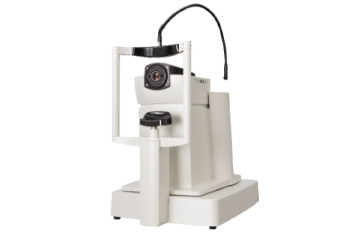
The i-Trace, a patented product from the American company Tracey, is a cutting-edge wavefront aberration analyzer that uses Ray-Tracing technology. It combines six measurement devices—wavefront aberration measurement, corneal topography, autorefractor, automated keratometer, pupil size measurement, and tear film analyzer—into a single all-in-one device. This remarkable equipment can perform dozens of measurements that would typically require six separate machines, all within just one minute.

<<i-trace>>

The only device using Ray Tracing for wavefront analysis. Utilizes a low-intensity (infrared, 785 nm wavelength) HeNe diode laser. Sequential measurement of 256 rays in various shades.

  • 256 rays are sequentially projected to various locations of the pupil.
  • Measures the deviation within the eye (the error distance between the expected arrival point on the retina and the actual arrival point).
  • 1) Quantifies cataract opacity to provide objective evaluation.
  • 2) Separates measurements of vision loss due to dry eyes and cataracts.
  • 3) Instead of merely matching simple numbers for visual acuity measurements, it reproduces the actual visual phenomena experienced by patients, effectively depicting the quality of vision through graphical representation (such as the “E” letter simulation).
  • 4) Quantifies the dysfunction of the lens caused by cataract (Dysfunctional Lens Index) and couples anatomical diagnosis of cataracts with functional diagnosis of vision loss, assisting in personalized surgical decision-making and outcome prediction.
  • 5) Quantifying the tear layer examination scores allows for predictions regarding progression and potential impacts on cataract surgery, enabling a rational decision on the timing of the procedure and whether to utilize monofocal or multifocal lenses.
  • 6) Measurements of coma and high-order aberrations, as well as low-order aberrations, provide critical data for assessing the suitability of premium cataract surgery (simultaneously correcting myopia, hyperopia, astigmatism, and presbyopia). The Alpha angle, which reflects the error between the visual axis and the intraocular lens axis, is evaluated alongside the determination of astigmatism axes that account for both corneal and intraocular astigmatism. Additionally, the light distribution patterns of multifocal lenses are analyzed. These comprehensive examination results are essential for selecting the most appropriate intraocular lens and guiding lens for optimal surgical outcomes.

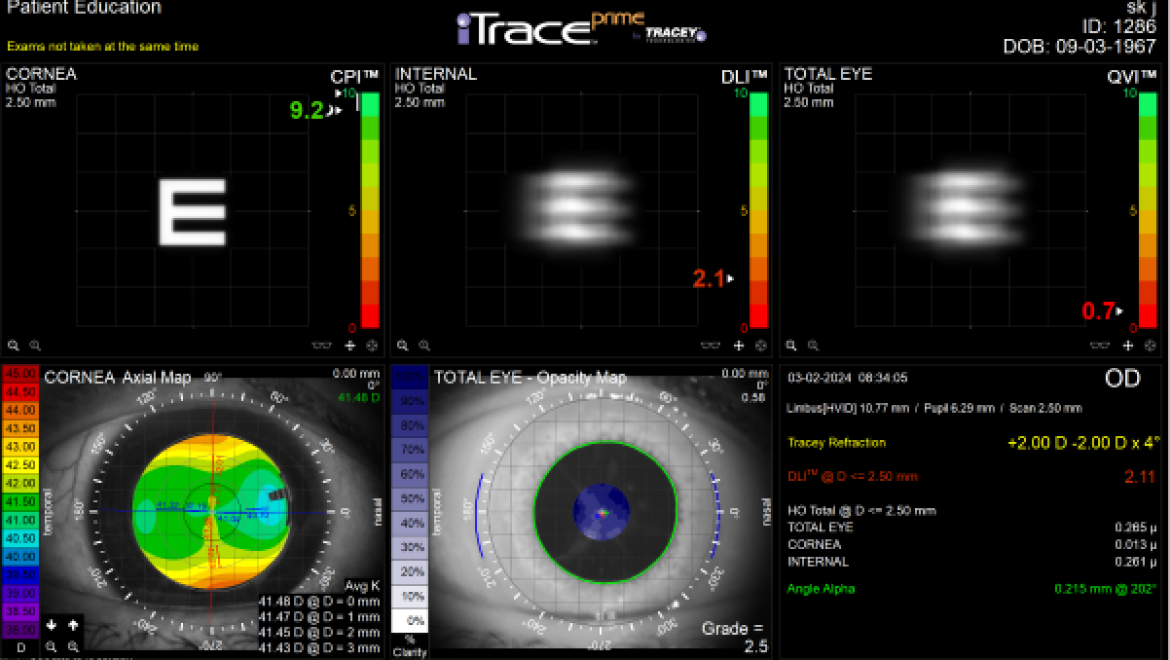
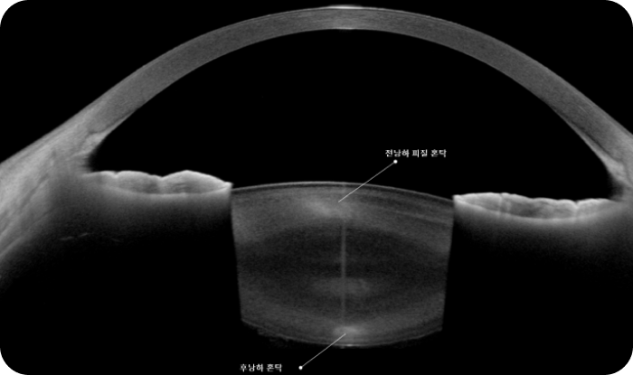
< Figure 1 & 2 - Cases of Central Cortical Opacity Measured by i-Trace Prime >

The patient underwent LASIK surgery 20 years ago, with unaided vision now measuring approximately 0.5. Routine examinations revealed corneal opacities alongside myopic regression and dry eye symptoms. The cataract opacity was small and not severe, leading to a treatment plan that included only artificial tears. According to the i-Trace differential examination, the corneal area (CORNEA) scored 9.2/10, while the lens area (INTERNAL) scored 2.1/10, indicating that light scattering caused by the lens was the primary reason for the vision decline. Following cataract surgery, the patient regained vision of 1.0. This case illustrates anatomical opacities present in multiple locations, with their severity not correlating directly to the visual acuity. The special feature of i-Trace, which allows for the separation of measurements from the cornea, lens, and retina to assess each visual function, played a crucial role in this assessment through the "light scattering optical characteristic analysis."

Cataract Symptoms

  • Vision Decrease

    Many patients experience double vision or glare during bright daylight. As cataracts progress, they may initially notice a slight decrease in distance vision, which gradually worsens. Vision becomes hazy, and myopia may worsen, making it difficult to see clearly even with previously suitable glasses or contact lenses

  • Pupil Opacity

    What appears to be a clear lens may gradually turn white, causing the normally black pupil to look white.

  • Monocular Diplopia

    Individuals who wear glasses or previously had good vision may see overlapping images when looking at objects with one eye.

  • Glare

    Bright environments can lead to significant glare, causing tearing and difficulty keeping the eyes open.

Cataracts typically progress slowly, making early symptoms subtle, and many patients may not notice changes until vision in the affected eye declines significantly. Those with cataracts in one eye may not realize the condition is progressing due to the unaffected eye's vision.

Additionally, some patients who previously needed reading glasses may suddenly find they can read without them due to the lens hardening and changing refractive index as cataracts develop. However, as cataracts progress further, opacity may increase to the point of total vision loss.

Cataract Prevention and Treatment

Prevention of Cataracts

Since cataracts result from oxidative stress-induced changes in lens proteins, wearing sunglasses, wide-brimmed hats, and using umbrellas to block UV rays, along with managing eye fatigue, can help prevent cataracts. Maintaining overall health by managing conditions like hypertension and diabetes, eating a balanced diet rich in vitamins E, B2, C from vegetables and fruits, and avoiding smoking can also delay the onset of cataracts.

Treatment of Cataracts

In the early stages of cataracts, treatment with eye drops (such as Garuni, Curen, and Posod) may be considered.

These eye drops can help slow the progression of cataracts by inhibiting the protein degeneration in the lens. However, the primary purpose of these drops is to "suppress the progression of cataracts," meaning they do not restore the cloudy areas of the lens to their original transparent state.

Therefore, it can be concluded that no current medication exists to treat cataracts effectively. The only viable treatment method for cataracts involves removing the cloudy lens and replacing it with a transparent intraocular lens (IOL).

While cataracts are not an urgent condition requiring immediate treatment, if they progress to the point of significantly impacting daily life and do not improve with medication, surgery should be considered

Types and Characteristics of
Intraocular Lenses Used in Cataract Surgery

Monofocal and Multifocal Lenses

Intraocular lenses used in cataract surgery include monofocal and multifocal IOLs.
The lenses covered by health insurance are monofocal IOLs, which allow good distance vision post-surgery, but require reading glasses for near tasks. Conversely, if the IOL is set for near vision, glasses will be necessary for distance.

  • Multifocal Lenses

  • Monofocal Lenses

Multifocal IOLs are more expensive but can correct myopia, hyperopia, astigmatism, and presbyopia, offering solutions for both near and distance vision as well as trifocal and quadrifocal options.

Types of Multifocal Intraocular Lenses

  • alcon
    Clareon Panoptix

    Awarded the prestigious Medical Technology Award by the American Preclinical Institute based on the technological prowess of AcriSof PanOptix.
    A multifocal IOL utilizing four focal points, providing clear vision for near, intermediate, and far distances with 88% light utilization.
    Aspheric design enhances night vision.

  • hoya
    Vivinex Gemetric

    Glare-free hydrophobic acrylic material.
    Proprietary aspheric optical design improves image quality.
    Reduces posterior capsule opacification through active oxygen treatment and square optic edge design.
    Excellent rotational stability for effective astigmatism correction.

  • zesis
    Lisa-Tri

    Trifocal design improves near, intermediate, and far vision.
    Minimizes light scattering due to diffraction, enhancing visual quality.
    Maximizes light transmission to the retina.
    Corrects astigmatism.

  • gohnron gohnron
    Tecnis Eyhance

    Provides enhanced intermediate vision compared to standard cataract surgery.
    Improves clarity at typical intermediate distances (approximately 66 cm).
    Minimizes glare with no diffraction rings or optical aberrations.
    Reduces light dispersion, decreasing discomfort during night driving.

Cataract Surgery Method

Cataract surgery involves creating a small opening in the black or white part of the eye, through which an ultrasound device is inserted to aspirate the cloudy lens caused by cataracts. A permanent artificial lens, similar in shape to the natural lens, is then inserted in its place. Just as eyeglass lenses come in various prescriptions to accommodate different visual needs, the power of the artificial lens is selected based on the patient's vision to ensure clear sight for either near or far distances.

  • step 1
  • step 2
  • step 3

Post-Surgery Care

For a week post-surgery, antibiotic and anti-inflammatory eye drops are used. During this time, the surgical site heals, and vision begins to recover. If the patient has other eye conditions, such as diabetic retinopathy, macular disease, glaucoma, or optic nerve damage in addition to cataracts, recovery may be delayed, or surgical outcomes may be unsatisfactory. It is advisable to avoid heavy physical labor, exercise, and activities that may allow dust or foreign objects to enter the eye for about a week after surgery. Watching TV, reading, using a computer, and using smartphones are acceptable. It is recommended to wear protective eye shields while sleeping for about three weeks post-surgery.

Progress

If cataracts do not interfere with daily life, it is fine to continue as is. However, if cataracts worsen, surgery should be considered. Cataract surgery removes the cloudy lens and replaces it with an artificial lens. If an artificial lens is not inserted, severe hyperopia will occur, necessitating the use of thick convex lenses. Therefore, unless in special cases, inserting an artificial lens is essential. Once implanted, an artificial lens typically does not need to be replaced for a lifetime, except in exceptional circumstances.

DR.ICL Eye Clinic conducts
thorough examinations to assess the patient's eye condition.

After the examination, personalized consultations
with medical staff determine the best treatment for presbyopia.

  • Precision Examination

    Before consulting with medical staff, a one-on-one precision examination with an optometrist assesses the patient's eye condition.

  • Medical Staff Consultation

    Based on the examination results, a one-on-one consultation with medical staff is conducted. At this time, the medical staff decides on the most suitable treatment method for the patient.

  • Surgery Progress

    The surgery is performed according to the treatment method decided during the medical consultation.

There are many types of cataract lenses available, and new lenses are continuously being developed.
Not all new lenses are necessarily better. In fact, their limited clinical experience may increase the likelihood of unexpected side effects. Simply opting for a new lens does not guarantee better outcomes; it is essential to choose a lens that suits the individual. Determining what works best for you starts with an accurate examination of your eyes. DR.ICL Eye Clinic leverages overwhelming surgical experience and advanced cataract diagnostic equipment—i-TRACE, ANTERION, CASIA2—to perform surgeries in 10 minutes using the most appropriate guiding lenses and the most comfortable artificial lenses, rather than just the most expensive ones.